Dermoscopy in Toowoomba
Dermoscopy and Skin Cancer Assessment in Toowoomba
Dermoscopy is an important part of modern skin cancer assessment. It allows a doctor to examine moles and skin lesions in greater detail than is possible with visual inspection alone.
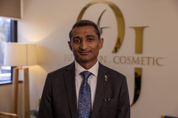
At OJ Clinics in East Toowoomba, dermoscopy is used as part of every full body skin check and focused mole assessment. Examinations are performed by Dr Omi Jindal, a specialist General Practitioner with postgraduate training in skin cancer medicine and dermoscopy from University of Queensland.
If a lesion requires further investigation, skin biopsies and many follow-up procedures can be performed at the clinic under local anaesthetic.
- Doctor-led skin examination.
- Dermoscopic mole assessment.
- Full body skin checks.
- Skin biopsies available on site.
- Digital dermoscopy.
- No referral required.
- Medicare rebates may apply.
What Is Dermoscopy?
Dermoscopy, sometimes called dermatoscopy, is a non-invasive technique used to examine moles and other skin lesions.
The examination is performed using a handheld instrument called a dermatoscope. It combines magnification with specialised lighting, allowing the doctor to assess colours, structures, blood vessels and pigment patterns beneath the outer surface of the skin.
Two lesions may look similar to the naked eye but appear very different when examined with a dermatoscope. One may show an orderly pattern consistent with a benign mole, while another may have irregular features that require monitoring, biopsy or removal.
Dermoscopy is painless and usually takes only a few seconds for each lesion. The instrument is placed lightly against the skin or held just above it.

Why Is Dermoscopy Important?
Some skin cancers are easy to recognise, while others can be subtle during their early stages.
Dermoscopy helps the doctor examine features that cannot be assessed properly through ordinary visual inspection. It may help distinguish between benign lesions and those that require further investigation.
Dermoscopic examination can assist with the assessment of:
- Melanoma.
- Basal cell carcinoma.
- Squamous cell carcinoma.
- Actinic keratoses.
- Benign moles.
- Seborrhoeic keratoses.
- Vascular lesions.
- Other pigmented and non-pigmented skin lesions.
Dermoscopy can also help avoid unnecessary procedures when a lesion has reassuring features. However, it is an examination aid rather than a definitive laboratory test.
No clinical examination can identify every skin cancer with complete certainty. When a lesion remains suspicious or cannot be confidently diagnosed, a biopsy may be recommended.
Dermoscopy at OJ Clinics
Dermoscopy is a routine part of skin cancer assessment at OJ Clinics.
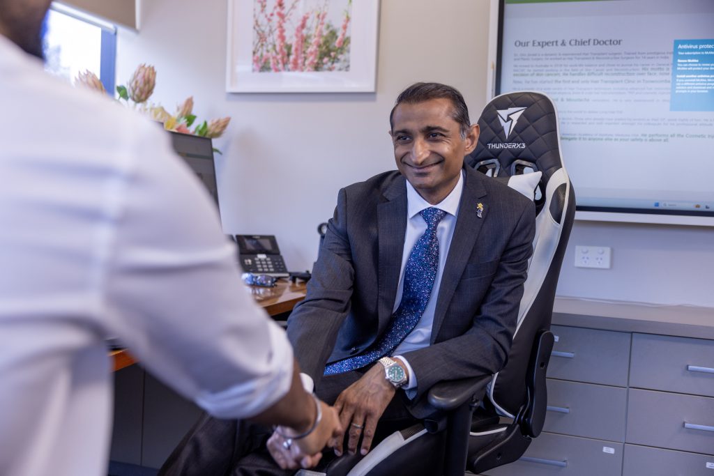
Your examination is performed by Dr Omi Jindal, a General Practitioner who:
- Holds a Master of Medicine in Skin Cancer Medicine from The University of Queensland.
- Has undertaken postgraduate training in dermoscopy and skin cancer assessment.
- Has an extensive background in general, plastic and reconstructive surgery.
Dr Jindal does not rely on visual inspection alone when assessing a concerning mole or skin lesion. Where appropriate, the lesion is examined with a dermatoscope and assessed in the context of your medical history, sun exposure and personal risk factors.
The findings are explained during the appointment so that you understand what has been seen and what should happen next.
What Happens During a Dermoscopic Examination?
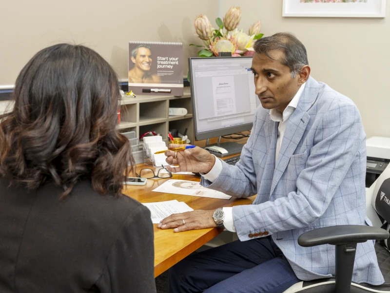
A dermoscopic assessment is straightforward and does not require needles, anaesthetic or removal of tissue.
- Medical and Skin History
Dr Jindal may ask about:
- How long the lesion has been present.
- Whether it has changed in size, shape or colour.
- Symptoms such as bleeding, itching or tenderness.
- Previous skin cancers or biopsies.
- Family history of melanoma.
- Occupational and recreational sun exposure.
- Previous severe sunburn.
- Current medications.
- Medical conditions affecting the immune system.
2. Visual Examination
The lesion is first assessed with the naked eye. Its size, shape, colour, surface and relationship to the surrounding skin are considered.
3. Dermoscopic Examination
The dermatoscope is placed gently against the skin or held close to the lesion.
Dr Jindal examines the lesion’s internal structures, including:
- Pigment patterns.
- Symmetry.
- Borders.
- Colours.
- Blood vessel patterns.
- Surface structures.
- Areas of regression or inflammation.
4. Discussion of Findings
The findings are explained in clear language.
Depending on the assessment, the next step may be:
- Reassurance.
- Self-monitoring.
- Clinical or dermoscopic photography.
- Review after a defined period.
- Skin biopsy.
- Complete removal of the lesion.
- Specialist referral.
Dermoscopy During a Full Body Skin Check
During a comprehensive skin check, Dr Jindal examines the skin systematically from head to toe.
Dermoscopy is used to assess lesions that require closer examination, including moles, sun-damaged areas and spots that appear unusual or different from the surrounding lesions.
A full body skin check may be more appropriate than a focused mole check if you:
- Have several lesions of concern.
- Have many moles.
- Have significant sun damage.
- Have previously had melanoma or another skin cancer.
- Have a family history of melanoma.
- Work or have worked outdoors.
- Have not previously had a professional skin examination.
A focused dermoscopic mole check may be suitable when you have one or two specific lesions that concern you.
Dermoscopy for Melanoma Assessment
Melanoma may develop within an existing mole or appear as a new lesion.
Some melanomas follow the familiar ABCDE pattern:
A — Asymmetry
B — Border irregularity
C — Colour variation
D — Diameter
E — Evolving or changing
However, not every melanoma follows these rules. Some may be small, evenly coloured, pink, red or almost colourless.
Dermoscopy allows the doctor to look for subtle patterns that may not be visible on the skin surface. A lesion that has suspicious dermoscopic features will usually require biopsy or removal for pathology testing.
Dermoscopic Monitoring
Not every mildly unusual mole needs to be removed immediately. In selected cases, careful monitoring may be appropriate.
The lesion may be documented or photographed so that its appearance can be compared at a future appointment. Monitoring can be useful when a lesion does not currently justify biopsy but should not be ignored.
You will be advised:
- Why monitoring is appropriate.
- When the lesion should be reviewed.
- Which changes should prompt an earlier appointment.
- Whether a biopsy may be required if the lesion evolves
Monitoring does not replace follow-up. You should return promptly if the lesion changes before the planned review date.
Patients with many moles or a higher risk of melanoma may also benefit from total body photography or more structured surveillance.
What If Dermoscopy Shows a Suspicious Lesion?
A suspicious dermoscopic appearance does not always mean that a lesion is cancerous. It means that further investigation is needed to establish the diagnosis.
Dr Jindal may recommend:
- A shave biopsy.
- A punch biopsy.
- An incisional biopsy.
- An excision biopsy.
- Complete surgical removal.
- Referral to a dermatologist or another specialist.
The biopsy method depends on the lesion’s size, depth, appearance, location and suspected diagnosis.
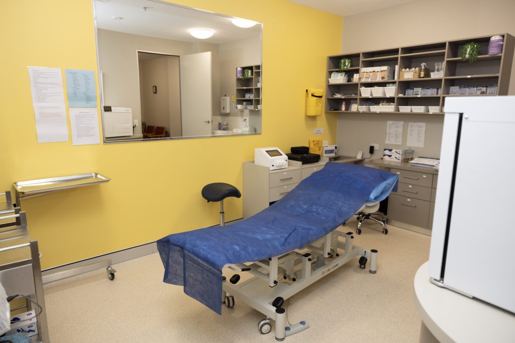
Most skin biopsies can be performed at OJ Clinics under local anaesthetic.
Skin Biopsy and Pathology
A skin biopsy involves removing a sample of tissue and sending it to a pathology laboratory for examination under a microscope.
Pathology can determine:
- The type of lesion.
- Whether skin cancer is present.
- The type and behaviour of the skin cancer.
- Whether further treatment is required.
- Whether the lesion appears to have been completely removed.
Once the result is available, Dr Jindal will explain the findings and discuss the next stage of care.
What If Skin Cancer Is Diagnosed?
Many skin cancers can be treated at OJ Clinics where this is clinically appropriate.
Treatment may include:
- Surgical excision.
- Curettage and cautery.
- Cryotherapy for selected lesions.
- Direct wound closure.
- Local flap reconstruction.
- Skin graft reconstruction.
- Wound care and postoperative review.
- Ongoing skin cancer surveillance.
Some lesions are better managed by a dermatologist, plastic surgeon, head and neck surgeon or hospital-based service.
If specialist care is required, Dr Jindal will explain why and arrange an appropriate referral. Where suitable, he will remain involved in reviewing reports, explaining the treatment pathway and coordinating your follow-up care.
Is Dermoscopy Accurate?
Dermoscopy provides more information than visual inspection alone, but it is not necessary that every skin cancer will be detected.
Its value depends on:
- The training and experience of the practitioner.
- The quality of the clinical examination.
- The appearance and stage of the lesion.
- The patient’s medical history and risk factors.
- Appropriate follow-up when monitoring is recommended.
When there is uncertainty, pathology testing remains the most reliable way to establish a diagnosis.
Does Dermoscopy Hurt?
No. Dermoscopy is painless and non-invasive.
The dermatoscope touches the skin lightly or is held just above it. No tissue is removed during the examination, and no radiation is involved.
If a biopsy or excision is recommended, that is a separate procedure performed under local anaesthetic.
How Should I Prepare for a Dermoscopic Skin Check?
No special preparation is required, but the following steps can make the examination easier:
- Avoid makeup where possible.
- Remove nail polish from fingers and toes.
- Avoid fake tan before the appointment.
- Avoid applying heavy creams or body oils on the day.
- Wear clothing that is easy to remove.
- Bring previous pathology reports if available.
- Make a note of any lesions that have changed.
- Bring photographs that show how a spot has evolved.
For a full body skin check, you will usually be asked to undress to your underwear. The examination is conducted in stages, and your privacy is respected throughout. You may request a chaperone.
Dermoscopy for Children and Teenagers
Children commonly develop new moles as they grow, and most are harmless.
Dermoscopy can be used to examine a concerning mole, birthmark or skin lesion without cutting the skin. It may be helpful when a lesion:
- Is growing quickly.
- Has changed in shape or colour.
- Bleeds or crusts.
- Becomes painful or itchy.
- Looks different from the child’s other moles.
- Appears as a persistent pink or red bump.
- Causes concern to a parent or guardian.
The examination is performed gently, with a parent or guardian present.
Most childhood moles do not require removal. Where further investigation is recommended, the reasons, options and risks will be explained clearly.
Why Choose OJ Clinics for Dermoscopy?
Doctor-Led Assessment
Your examination is performed by Dr Omi Jindal, a General Practitioner with postgraduate qualifications in skin cancer medicine.
Dermoscopy as Standard Practice
Dermoscopy is included as part of full body skin checks and focused mole assessments where clinically appropriate.
On-Site Biopsy
Many skin biopsies and minor procedures can be performed at the clinic under local anaesthetic.
Pathology and Follow-Up
Pathology results, treatment planning, wound care and ongoing skin surveillance can be coordinated through the same clinic.
Surgical and Reconstructive Experience
Where excision is required, Dr Jindal’s reconstructive background supports careful planning of wound closure, local skin flaps and skin grafts.
Referral When Required
Specialist or hospital-based care is arranged when it offers a safer or more appropriate treatment pathway.
No Referral Required
You can contact OJ Clinics directly to arrange a dermoscopic skin check or mole assessment.
Dermoscopy in East Toowoomba and the Darling Downs
OJ Clinics provides dermoscopic skin checks and mole assessments for patients from Toowoomba and surrounding communities, including:
- East Toowoomba.
- South Toowoomba.
- North Toowoomba.
- Rangeville.
- Middle Ridge.
- Kearneys Spring.
- Harristown.
- Glenvale.
- Highfields.
- Drayton.
- Oakey.
- Pittsworth.
- The wider Darling Downs region.
We are committed to maintaining the highest standards of patient privacy. We never use or publish your photographs or personal information.
Other Services at OJ Clinics
Frequently Asked Questions
A skin check is the overall examination of your skin. Dermoscopy is the specialised technique used to examine individual moles and lesions in greater detail.
Dermoscopy is used where clinically appropriate, particularly for pigmented, vascular or otherwise unusual lesions.
Dermoscopy can identify suspicious features, but a definitive melanoma diagnosis generally requires pathology examination of a biopsy or excised lesion.
In some cases, a biopsy can be performed during the same visit. A separate procedure appointment may be needed depending on the lesion, available time and type of biopsy required.
Yes. Dermoscopy can assist with the assessment of both pigmented and non-pigmented lesions, including some basal cell carcinomas and squamous cell carcinomas.
Yes. Dermoscopy is non-invasive and can be used to assess moles and other skin lesions in children and teenagers.
No. You can book a dermoscopic skin check or mole assessment directly with OJ Clinics.
Medicare rebates may apply to eligible medical consultations and procedures. Confirm expected fees and out-of-pocket costs with the clinic when booking.
Book a Dermoscopic Skin Check in Toowoomba
Book an appointment if you have noticed:
- A new mole
- A changing spot
- A lesion that looks different from your other moles
- A persistent sore
- A spot that bleeds or crusts
- A lesion that has returned after previous treatment