Total Body Photography
Total Body Photography & Mole Mapping in Toowoomba
If you have many moles, a previous melanoma or other significant risk factors for skin cancer, a single skin check may not provide enough information to monitor your skin effectively over time.
Structured skin surveillance creates a clear record of your skin and helps identify lesions that are new or changing. Depending on your individual risk, this may include regular full body skin checks, dermoscopic assessment, monitoring of selected lesions and formal total body photography, commonly known as mole mapping.
At OJ Clinics in East Toowoomba, Dr Omi Jindal provides GP-led skin cancer surveillance and helps determine which level of monitoring is appropriate for you. Where formal total body photography is recommended, referral can be arranged to a suitable imaging service, with your ongoing skin checks and follow-up coordinated through OJ Clinics.
No referral is required to book a skin surveillance consultation.
- GP-led skin cancer surveillance.
- Comprehensive full body skin checks.
- Dermoscopic examination.
- Monitoring of selected moles.
- Referral for formal mole mapping.
- Skin biopsy available on site.
- No referral required.
What Is Total Body Photography?
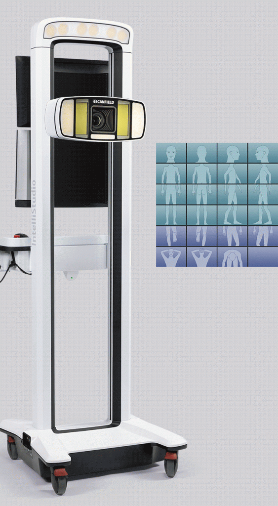
Total body photography is a method of recording the appearance and distribution of moles and other skin lesions across the body.
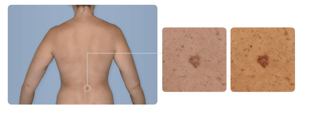
A series of standardised photographs is taken to create a baseline record of the skin. At later appointments, new images can be compared with the original photographs to identify spots that have appeared or changed.
Total body photography is also referred to as:
- Mole mapping.
- Full body mole photography.
- Skin mapping.
- Photographic skin surveillance.
The photographs provide an objective reference that can be particularly useful for people with many moles or lesions that are difficult to monitor without imaging.
How Does Mole Mapping Help?
Changes in a mole may occur gradually and can be difficult to recognise from memory alone.
Comparing current photographs with previous images may help identify:
- A new lesion.
- An increase in size.
- A change in shape or symmetry.
- New or changing pigmentation.
- Alteration in the surface or elevation of a mole.
- A lesion that now looks different from surrounding spots.
Change over time is an important warning sign when assessing melanoma. Some early melanomas may not initially look obviously abnormal but become concerning because they are new or evolving.
Mole mapping does not diagnose melanoma. Any new or changing lesion still requires examination by a doctor, usually with dermoscopy. A biopsy may be needed to establish a definite diagnosis.
Total Body Photography, Dermoscopy and Mole Monitoring
These methods have different roles and are often used together.
Total Body Photography
Total body photography records the overall pattern of moles and lesions across the skin. It is primarily used to identify new or visibly changing spots.
Dermoscopy
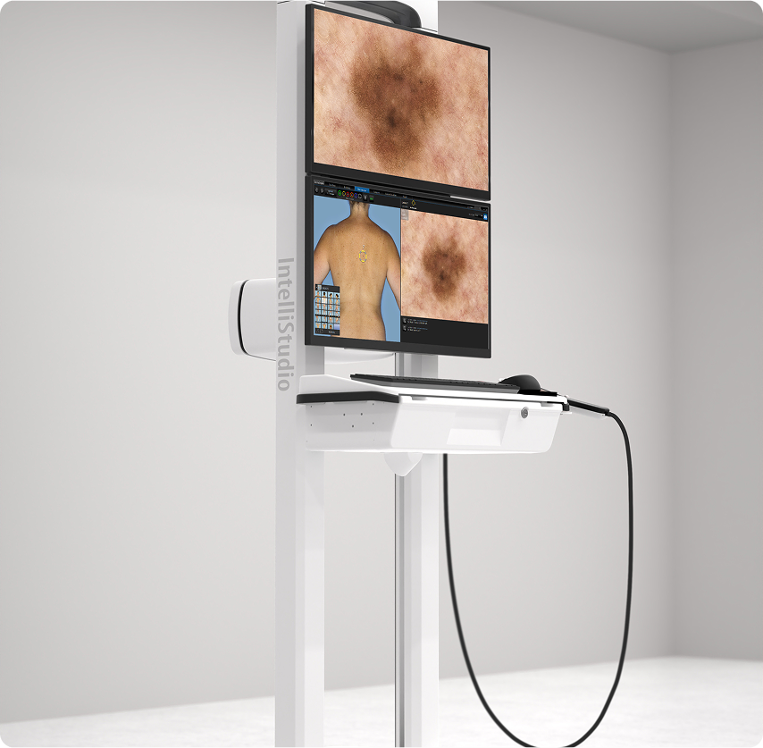
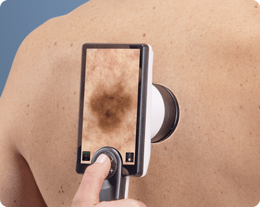
Dermoscopy provides a magnified view of an individual lesion. It allows the doctor to assess pigment, blood vessels, structures and patterns beneath the skin surface that are not visible during an ordinary visual examination.
Dermoscopic Lesion Monitoring
Selected moles may be documented and reviewed over time when immediate biopsy is not considered necessary. This allows the doctor to look for structural changes at follow-up appointments.
The most appropriate combination depends on your melanoma risk, mole count, examination findings and previous skin cancer history.
Who May Benefit From Mole Mapping?
Formal total body photography may be helpful for people at increased risk of melanoma, particularly those with:
- A personal history of melanoma.
- A strong family history of melanoma.
- A large number of moles.
- Multiple atypical or irregular-looking moles.
- Moles that are difficult to monitor at home.
- A history of repeatedly changing lesions.
- Significant sun damage.
- Fair skin that burns easily.
- A history of severe or blistering sunburn.
- A weakened immune system.
- A genetic condition associated with increased melanoma risk.
- Difficulty determining whether a lesion is new.
The presence of one risk factor does not automatically mean that mole mapping is required. A medical assessment is the best way to determine whether it is likely to provide meaningful benefit.
Does Everyone Need Total Body Photography?
No. Total body photography is not required for every person who has moles.
For many people, an appropriate surveillance plan may consist of:
- Regular self-examination
- A professional full body skin check
- Dermoscopic assessment of suspicious lesions
- Follow-up at an interval based on individual risk
Formal mole mapping is generally more useful for patients with numerous moles, previous melanoma or other significant risk factors.
Dr Jindal will assess your circumstances and explain whether total body photography is likely to add value to your ongoing care.
Skin Surveillance at OJ Clinics
Skin surveillance at OJ Clinics is based on your personal risk rather than a standard schedule applied to every patient.
Your plan may include the following.
Skin Cancer Risk Assessment
Dr Jindal will review factors that may affect your risk, including:
- Previous melanoma or other skin cancers
- Previous biopsies and pathology results
- Family history of melanoma
- Number and pattern of moles
- Skin type
- Degree of sun damage
- Occupational and recreational sun exposure
- Previous severe sunburn
- Medications that affect the immune system
- Your ability to examine your own skin
This assessment helps determine how often you should be reviewed and whether additional imaging is appropriate.
Comprehensive Skin Examination
A systematic examination of the skin is performed to identify new, changing or suspicious lesions.
The examination may include:
- Scalp and hairline
- Face, ears and neck
- Chest, abdomen and back
- Arms, hands and fingers
- Legs, feet and toes
- Soles of the feet
- Nails and surrounding skin
Your privacy and comfort are respected throughout the consultation.
Dermoscopic Assessment
Moles and other lesions of interest are examined with a dermatoscope where appropriate.
Dermoscopy can help determine whether a lesion:
- Appears reassuring
- Should be monitored
- Requires biopsy or removal
- Needs specialist assessment
Monitoring Selected Moles
Some lesions may not require immediate removal but should not simply be ignored.
Where monitoring is appropriate, the lesion’s clinical and dermoscopic features can be documented and a review interval recommended. You should return earlier if the lesion changes before the scheduled appointment.
Planned Follow-Up
The frequency of surveillance varies from person to person.
Patients with a previous melanoma, multiple atypical moles or significant skin cancer risk may require more frequent review. Others may only need periodic skin checks or assessment when a new concern arises.
No Referral needed for Formal Total Body Photography
OJ Clinics provides medical skin surveillance, including full body skin checks and dermoscopic examination.
Where formal total body photography is likely to improve monitoring, Dr Jindal can arrange referral to an appropriate imaging provider.
Referral may be considered if you have:
- A previous melanoma and numerous moles
- A very high mole count
- Multiple atypical naevi
- Lesions that are difficult to monitor without photographs
- A risk profile requiring more structured long-term surveillance
The resulting images can then support future clinical skin examinations.
Dr Jindal can remain involved in reviewing your skin, assessing lesions identified through imaging, arranging biopsies where required and coordinating follow-up care.
What Happens at a Skin Surveillance Consultation?
Your first consultation generally includes:
Medical History
You will be asked about previous skin cancers, biopsies, family history, sun exposure and any lesions that concern you.
Risk Assessment
Your personal melanoma and skin cancer risk will be considered.
Full Body Skin Check
Your skin will be examined systematically for new, changing or unusual lesions.
Dermoscopic Examination
Selected moles and skin lesions will be examined under magnification using a dermatoscope.
Personalised Surveillance Plan
Dr Jindal will explain whether you may benefit from:
- Routine skin checks
- More frequent surveillance
- Monitoring of selected lesions
- Formal total body photography
- Biopsy or excision
- Specialist referral
The reasons for each recommendation will be discussed with you.
What Happens If a Suspicious Lesion Is Found?
A suspicious lesion does not necessarily mean that skin cancer is present.
Depending on the clinical and dermoscopic findings, Dr Jindal may recommend:
- Reassurance
- Monitoring at home
- Review after a defined period
- Clinical or dermoscopic photography
- Skin biopsy
- Complete removal of the lesion
- Referral to a dermatologist or another specialist
When a biopsy is performed, the tissue is sent to a pathology laboratory for examination under a microscope.
Skin Biopsy and Treatment
Many skin biopsies and selected skin cancer procedures can be performed at OJ Clinics under local anaesthetic.
Procedures may include:
- Shave biopsy
- Punch biopsy
- Incisional biopsy
- Excision biopsy
- Surgical excision
- Direct wound closure
- Local flap reconstruction
- Skin graft reconstruction
The recommended procedure depends on the lesion’s appearance, size, depth, location and suspected diagnosis.
Biopsies and excisions are invasive procedures and carry risks, including bleeding, infection, delayed healing and scarring. The relevant risks, expected recovery and treatment alternatives will be discussed before you decide whether to proceed.
Why Ongoing Skin Surveillance Matters
A skin check records what your skin looks like at one point in time. Surveillance allows your skin to be assessed for change.
An ongoing plan may involve:
- Becoming familiar with your own skin
- Looking for new or changing lesions
- Attending scheduled medical reviews
- Comparing lesions over time
- Investigating suspicious changes promptly
- Adjusting the review interval as your risk changes
Some people require close surveillance, while others may only need periodic examination.
The aim is to provide enough monitoring to identify meaningful change without performing unnecessary procedures.
Checking Your Skin Between Appointments
Continue to examine your skin between professional reviews.
Arrange an earlier appointment if you notice a lesion that:
- Is new
- Changes in size, shape or colour
- Looks different from your other moles
- Bleeds or crusts repeatedly
- Becomes painful, itchy or tender
- Develops several colours
- Does not heal
- Returns after previous treatment
Do not wait for your next scheduled skin check if you notice a concerning change.
The ABCDE Guide
The ABCDE guide can help identify some suspicious moles:
- A — Asymmetry: one half does not match the other
- B — Border: irregular, blurred or poorly defined edges
- C — Colour: several colours or a change in pigmentation
- D — Diameter: a larger lesion may require closer assessment
- E — Evolving: any change in size, shape, colour, surface or symptoms
Not every melanoma follows the ABCDE pattern. Some may be small, evenly coloured, pink, red or otherwise subtle.
Any new or changing lesion should be assessed even when it does not fit the usual warning signs.
Why Choose OJ Clinics for Skin Surveillance?
GP-Led Care
Your surveillance is managed by Dr Omi Jindal, a General Practitioner with postgraduate training in skin cancer medicine and dermoscopy.
Individualised Monitoring
The type and frequency of surveillance are based on your personal risk factors and examination findings.
Dermoscopic Examination
Moles and lesions of concern are examined with dermoscopy where appropriate.
On-Site Biopsy
Many skin biopsies and minor procedures can be performed at the clinic under local anaesthetic.
Referral for Formal Mole Mapping
Where total body photography is likely to be beneficial, referral can be arranged to an appropriate imaging provider.
Continuity of Care
Skin checks, imaging findings, biopsies, pathology results, treatment and future surveillance can be coordinated through the same clinic.
No Referral Required
You can book directly with OJ Clinics.
Serving Toowoomba and the Darling Downs
OJ Clinics provides skin cancer risk assessment and ongoing skin surveillance for patients from Toowoomba and surrounding areas, including:
- East Toowoomba
- Rangeville
- Middle Ridge
- Kearneys Spring
- South Toowoomba
- North Toowoomba
- Harristown
- Glenvale
- Highfields
- Drayton
- Oakey
- Pittsworth
- The wider Darling Downs region
We are committed to maintaining the highest standards of patient privacy. We never use or publish your photographs or personal information.
Other Services at OJ Clinics
Frequently Asked Questions
Mole mapping uses standardised photographs to create a baseline record of the moles and lesions on your skin. Future images can be compared with the baseline to identify new or changing spots.
No. Mole mapping provides photographic records, while a skin check is a medical examination performed by a doctor. Photography supports clinical examination but does not replace it.
No. Total body photography can help identify a lesion that is new or changing. The lesion must still be medically assessed, and a biopsy may be required for diagnosis.
Not necessarily. The benefit depends on your overall risk, the number and type of moles you have and whether they can be monitored reliably through regular skin checks.
Dermoscopic monitoring involves documenting the detailed appearance of a selected lesion and reassessing it at a future appointment for structural change.
The interval varies according to your risk profile, previous findings and the advice of your doctor or imaging provider.
The lesion should be examined by a doctor. Depending on its appearance, dermoscopic review, biopsy, removal or specialist referral may be recommended.
No referral is required to book a skin surveillance consultation at OJ Clinics. Dr Jindal can arrange a referral if formal imaging or specialist care is recommended.
Medicare rebates may apply to eligible medical consultations and procedures. Formal photography services may involve separate fees that are not covered, or are only partly covered, by Medicare or private health insurance. Costs should be confirmed with the imaging provider.
Book a Skin Surveillance Consultation in Toowoomba
Consider a structured skin surveillance plan if you have many moles, a previous melanoma, a strong family history of melanoma or other significant skin cancer risk factors.
Dr Jindal can assess your risk and advise whether you require regular skin checks, dermoscopic monitoring or referral for formal total body photography.